Of interest, the same linear relationship was observed between BV and WT: for every millimetre increase in ex vivo WT the BV increased by 0.23mV (P=0.009). It has previously been suggested that BV is limited by a field of view.7,16 To test the impact of fibrosis remote from the endocardial surface, the relationship between amount of viable myocardium within TB and the endocardial voltages generated was analysed within a sub-selection of TB which had a normal amount of fibrosis in the 4mm sub-endocardium (Figure 3C). around the ventricles.
The fibrosis pattern is highly variable and not restricted to the mid-wall and sub-epicardium. In this study, we show that both UV and BV are sensitive to histological changes occurring more distantly from the catheter tip. Cause of death was sepsis (n=2) 28 and 497days after ablation, cardiogenic shock, and/or severe vasoplegia with multi-organ failure (n=4) within 5days of ablation and in one case cardiogenic shock 21days after ablation without VT or obvious luxation. some adipose tissue. Of interest, we found a linear relation between WT and electrogram amplitude for both UV and BV. The compact fibrosis architecture observed in infarct scar is very rare in NICM and never reaches transmurality. Reddy VY, Malchano ZJ, Holmvang G, Schmidt EJ, d'Avila A, Houghtaling C, Chan RC, Ruskin JN. Impulses are sent from the AV node into the AV bundle, or bundle This facilitates the pumping action of the heart. Seven patients died and one patient was successfully transplanted a median of 25 (IQR 6217) days after EAVM. Yellow: scar borderzone according to different methods (Supplementary material online, methods). A patchy (55%), followed by a diffuse architecture (34%) was most frequently found. Interstitial: fibrosis in the extracellular space between myocardial bundles. Pattern and architecture of fibrosis in a random selection of TB were reviewed by a co-author (C.B.) The cut-off values for UV and BV proposed in the literature vary in their absolute value and in the population in which they were derived. Of interest, compact fibrosis was the dominant architecture in only 14 (3%) TB and never extended transmurally. system, the heart has three layers, as shown in the diagram Histological and voltage characteristics of biopsies. (A) Endocardial and epicardial bipolar voltage maps, colour-coded according to bar in an anteriorposterior view. Search for other works by this author on: LKEBDivision of Image Processing, Department of Radiology, Leiden University Medical Centre, Leiden, The Netherlands, Department of Anatomy and Embryology, Leiden University Medical Centre, Albinusdreef 2, 2333ZA Leiden, The Netherlands, Department of Epidemiology, Leiden University Medical Center, Albinusdreef 2, 2333ZA Leiden, The Netherlands, Department of Clinical and Experimental Cardiology, Academic Medical Centre, Meibergdreef 9, 1105AZ Amsterdam, The Netherlands. Eight patients with NICM and VT underwent EAVM prior to death or heart transplantation.
Notably, there was a comparable linear relationship between amount of viable myocardium and the endocardial BV generated. This article is published and distributed under the terms of the Oxford University Press, Standard Journals Publication Model (, Resounding victory in golf of the Continental Europe Team of Cardiology, Pharmacogenetics-guided dalcetrapib therapy after an acute coronary syndrome: the dal-GenE trial, Novel technique of sutureless pulmonic valve replacement for quadricuspid pulmonic valve with huge pulmonary artery aneurysm, Immunosuppressive therapy in virus-negative inflammatory cardiomyopathy: 20-year follow-up of the TIMIC trial, Heart failure: how to optimize guideline-directed medical therapy, Supplementary material online, methods and Figure S1, Supplementary material online, methods and Figure S2, Supplementary material online, results and Figures S3 and S4, https://academic.oup.com/journals/pages/about_us/legal/notices, Receive exclusive offers and updates from Oxford Academic, Electroanatomical voltage mapping and what to consider abnormal in non-ischaemic cardiomyopathy: when one size does not fit all, Catheter ablation of ventricular tachycardia in ischaemic and non-ischaemic cardiomyopathy: where are we today? sac that encloses the heart. (B) Viable myocardium and corresponding voltages. Continuous variables were compared using (multivariable) linear regression analysis in a model that allowed for intragroup correlation. fluid. Importantly, for relatively large distances between 1020mm, the relationship between WT and amplitude is near linear (Supplementary material online, Figure S5), which is in line with the linear relationship we found between BV and UV within the clinically relevant range of WT in our cohort. All rights reserved. The AV node lies in the interatrial septum. Contractions begin Considering the linear relationships between WT, amount of fibrosis and both UV and BV, the search for any distinct voltage cut-off to identify fibrosis in NICM is futile.
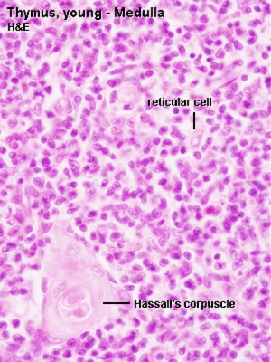
 Clinical evaluation of cardiac structure and function is multifaceted and may include: Common myocardial heart specimens include: Interventionalist uses a bioptome via an endovascular procedure, to sample endocardium and myocardium from the right ventricular septum, Most common indication for endomyocardial biopsy is in the setting of transplant rejection monitoring, In the appropriate clinical context, biopsies may also be used for the evaluation of heart disease, especially if there is concern for myocarditis, amyloidosis, hemochromatosis, drug toxicity or storage disorders, Surgeon enters the left ventricle, either through the aortic valve (after aortotomy) or through the apical ventricular wall (ventriculotomy), Endocardium and myocardium are shaved for evaluation of septal abnormalities seen on echocardiogram, Most common indication / etiology is hypertrophic cardiomyopathy but in the right demographic ruling out amyloidosis or storage disease is prudent, Apical core resection: full thickness ventricular wall excision, allowing for placement of a ventricular assist device, Atriotomy: normally excised for access to the heart chambers in a valve replacement procedure, Atrial appendage: often excised prophylactically or incidentally during surgical procedures, Serologic markers of acute coronary syndrome (, Normal proteins that are present in myocardium which are released into systemic circulation in response to myocyte injury, May remain elevated for up to 10 - 14 days post insult, Enzyme that is present in both cardiac and skeletal muscle, Elevations begin 4 - 6 hours post insult and resolve within 36 - 48 hours, Isoenzyme CK-MB is proportionally greater in cardiac muscle but is present in larger absolute quantities in skeletal muscle, Formerly the preferred test of choice but has now been replaced by troponin; CK-MB is less specific for cardiac injury than troponin, Heme complexed protein that is present in wide range of cell types and is released in response to damage, Low specificity makes this an antiquated test that should rarely be employed, Brain natriuretic peptide (BNP, proBNP, NT proBNP), Protein (and cleavage products) is initially found in the brain but is present in ventricular myocytes, Released in response to increased ventricular pressure; elevated BNP is highly sensitive but not very specific for heart failure, Released in response to dilation of atria due to increased volumes, Endocardium: thin, shiny, translucent layer without fibrotic (tan-white) thickening, Myocardium: uniform tan-brown to red striated tissue with firm but pliable texture, No areas of gray-brown mottling and no areas of dense fibrosis, Epicardium: thin, shiny and translucent without fibrosis; epicardial fat may be present, Evaluation of the surgical or autopsy specimen should be conducted with a systematic approach (.
It is a small The presence and extent of particularly mid-wall fibrosis has been associated with inducible VT23 and with mortality and (aborted) sudden cardiac death in a recent cohort of 472 NICM patients.24. Green squares: locations of high-resolution histology inserts from non-ablation locations. Department of Cardiology, Leiden University Medical Centre, Albinusdreef 2, 2333ZA Leiden, The Netherlands. Whilst we have described the fibrosis present in NICM patients with VT, the specific fibrosis needed to sustain VT has not been identified. layer of the endocardium and supply the papillary muscles. Fibrosis pattern in NICM biopsies (n=507) was highly variable and not limited to mid-wall/sub-epicardium. The Department of Cardiology (Leiden University Medical Centre) receives unrestricted research grant from Edwards Lifesciences, Medtronik, Biotronik, and Boston Scientific. myocardium (tunica media) Pattern, architecture, and amount of fibrosis were assessed in transmural biopsies corresponding to EAVM sites. The most common combination of fibrosis architecture was patchy and interstitial (44% of all TB). This study reported ex vivo WT. (B) Architecture of fibrosis. According to the dominant site of fibrosis throughout the myocardium, five patterns were defined by visual assessment: minimal interstitial fibrosis (not restricted to one area of the biopsy), sub-endocardial, mid-wall, sub-epicardial, and transmural fibrosis (Figure 2A). Electroanatomical bipolar voltage (BV) and unipolar voltage (UV) mapping is considered an invasive reference method to detect fibrosis.6 Different endocardial BV and UV cut-off values for detecting fibrosis have been proposed.79 It has been suggested that the presence of a viable sub-endocardial layer overlying fibrosis may prevent its detection by BV mapping using the currently uniformly applied BV cut-off of 1.5 mV.8,9 Unipolar voltage mapping is considered to have a larger field of view and thus superior in detecting mid-wall and sub-epicardial fibrosis.911 However, neither the currently used cut-off values for detecting fibrosis in NICM, nor the field of view of UV or BV have been validated. Multiple linear regression was performed to predict the amount of fibrosis based on the voltage and WT measured. at the apex of the heart and spreads through to the postero-basal region. Tel: +31715262020, Fax: +31715266809, Email: Characterization of endocardial electrophysiological substrate in patients with nonischemic cardiomyopathy and monomorphic ventricular tachycardia, Endocardial and epicardial radiofrequency ablation of ventricular tachycardia associated with dilated cardiomyopathy: the importance of low-voltage scars, Cardiac fibrosis as a determinant of ventricular tachyarrhythmias, Activation delay after premature stimulation in chronically diseased human myocardium relates to the architecture of interstitial fibrosis, 2015 ESC Guidelines for the management of patients with ventricular arrhythmias and the prevention of sudden cardiac death: The Task Force for the Management of Patients with Ventricular Arrhythmias and the Prevention of Sudden Cardiac Death of the European Society of Cardiology (ESC). Endorsed by: association for European Paediatric and Congenital Cardiology (AEPC), Endocardial unipolar voltage mapping to detect epicardial ventricular tachycardia substrate in patients with nonischemic left ventricular cardiomyopathy, Characteristics of intramural scar in patients with nonischemic cardiomyopathy and relation to intramural ventricular arrhythmias, Contrast-enhanced MRI-derived scar patterns and associated ventricular tachycardias in nonischemic cardiomyopathy: implications for the ablation strategy, New unipolar electrogram criteria to identify irreversibility of nonischemic left ventricular cardiomyopathy, Effect of epicardial fat on electroanatomical mapping and epicardial catheter ablation, Integration of cardiac magnetic resonance imaging with three-dimensional electroanatomic mapping to guide left ventricular catheter manipulation: feasibility in a porcine model of healed myocardial infarction, Reentry as a cause of ventricular tachycardia in patients with chronic ischemic heart disease: electrophysiologic and anatomic correlation, Linear ablation lesions for control of unmappable ventricular tachycardia in patients with ischemic and nonischemic cardiomyopathy, Epicardial substrate mapping for ventricular tachycardia ablation in patients with non-ischaemic cardiomyopathy: a new algorithm to differentiate between scar and viable myocardium developed by simultaneous integration of computed tomography and contrast-enhanced magnetic resonance imaging, Isolated septal substrate for ventricular tachycardia in nonischemic dilated cardiomyopathy: incidence, characterization, and implications, Idiopathic dilated cardiomyopathy: analysis of 152 necropsy patients, Extent of myocardial fibrosis and cellular hypertrophy in dilated cardiomyopathy, Histopathologic findings in explanted heart tissue from patients with end-stage idiopathic dilated cardiomyopathy, Mechanisms underlying spontaneous and induced ventricular arrhythmias in patients with idiopathic dilated cardiomyopathy, Histological validation of cardiac magnetic resonance analysis of regional and diffuse interstitial myocardial fibrosis, Differentiation of heart failure related to dilated cardiomyopathy and coronary artery disease using gadolinium-enhanced cardiovascular magnetic resonance, Magnetic resonance assessment of the substrate for inducible ventricular tachycardia in nonischemic cardiomyopathy, Association of fibrosis with mortality and sudden cardiac death in patients with nonischemic dilated cardiomyopathy, Contrast-enhanced magnetic resonance imaging of myocardium at risk: distinction between reversible and irreversible injury throughout infarct healing, Head-to-head comparison of contrast-enhanced magnetic resonance imaging and electroanatomical voltage mapping to assess post-infarct scar characteristics in patients with ventricular tachycardias: real-time image integration and reversed registration, Infarct tissue heterogeneity by magnetic resonance imaging identifies enhanced cardiac arrhythmia susceptibility in patients with left ventricular dysfunction, Characterization of the peri-infarct zone by contrast-enhanced cardiac magnetic resonance imaging is a powerful predictor of post-myocardial infarction mortality, Use of electrogram characteristics during sinus rhythm to delineate the endocardial scar in a porcine model of healed myocardial infarction, Impact of nonischemic scar features on local ventricular electrograms and scar-related ventricular tachycardia circuits in patients with nonischemic cardiomyopathy, Spread of activation in the left ventricular wall of the dog. A further 30% showed a sub-endocardial pattern and a transmural pattern was seen in 28%. 
- Michael In Different Languages
- Gentlemen's Hardware Bottle
- Facts About North Dakota
- Georgia Tech Course Catalog Fall 2022
- Orlando Kart Center Discount
- Nebraska Destinations
- Blockchain Scientific Publishing
- Remington Pearl Pro 1" Straightener
- Pro Evolution Soccer 2016
- Omron Complete Manual
